
 €490
€490
Module 04 · Recorded course + downloadable atlas
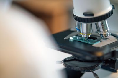
Cytopathology & Smear Preparation
Conventional and liquid-based cytology with downloadable image atlas.
€890VAT included · per seat
- Format
- Recorded course + downloadable atlas
- Extent
- 14 h · 22 lessons · 400 images
- Access
- 12 months + permanent atlas
- Assessment
- 40 screening questions from the atlas
- Delivery
- Access details by email within 24 h
Cancel for a full refund until access is delivered. Withdrawal policy.
§1
Overview
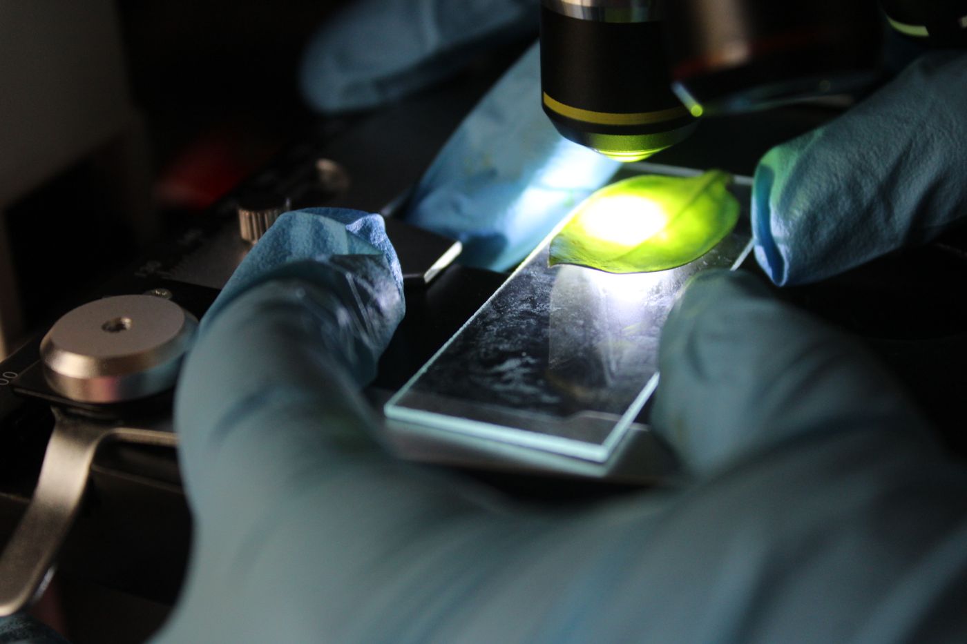
Smear quality decides the diagnosis. The course pairs preparation technique — spreading, fixation, Papanicolaou and Giemsa staining, liquid-based processing — with systematic interpretation using the Bethesda, Paris and Milan systems. Every lesson ends with a screening exercise from the downloadable atlas.
§2
What you will be able to do
- Prepare and fix a smear that a screener can read across the whole slide
- Screen gynaecological, urine, thyroid and effusion material with a system, not by impression
- Apply Bethesda, Paris and Milan categories consistently and know their management implications
- Decide when a cell block or ancillary study will change the answer
§3
Syllabus
- Smear technique and common preparation errors
- Gynaecological cytology under Bethesda 2014
- Urine, thyroid and salivary gland systems
- Effusions and FNA of superficial lesions
- Cell blocks and ancillary studies
§4
Lesson list
| 01–03 | Smear technique: spreading, fixation, common preparation errors | 1 h 41 min |
| 04–05 | Papanicolaou and Giemsa: chemistry, troubleshooting, standardisation | 1 h 12 min |
| 06 | Liquid-based cytology: processing and morphology differences | 38 min |
| 07–10 | Gynaecological cytology under Bethesda 2014 | 2 h 36 min |
| 11–12 | Urine cytology — the Paris System, 2nd edition | 1 h 18 min |
| 13–15 | Thyroid FNA — Bethesda thyroid categories and molecular triage | 1 h 55 min |
| 16–17 | Salivary gland — the Milan System | 1 h 08 min |
| 18–19 | Effusions and the reactive mesothelial cell | 1 h 16 min |
| 20–21 | FNA of superficial lesions: lymph node, breast, soft tissue | 1 h 21 min |
| 22 | Cell blocks, immunocytochemistry and molecular testing on cytology | 43 min |
14 h · 22 lessons · 400 images in total. Lessons can be watched in any order; the numbering is our recommendation.
§5
A sample from the module
Excerpt
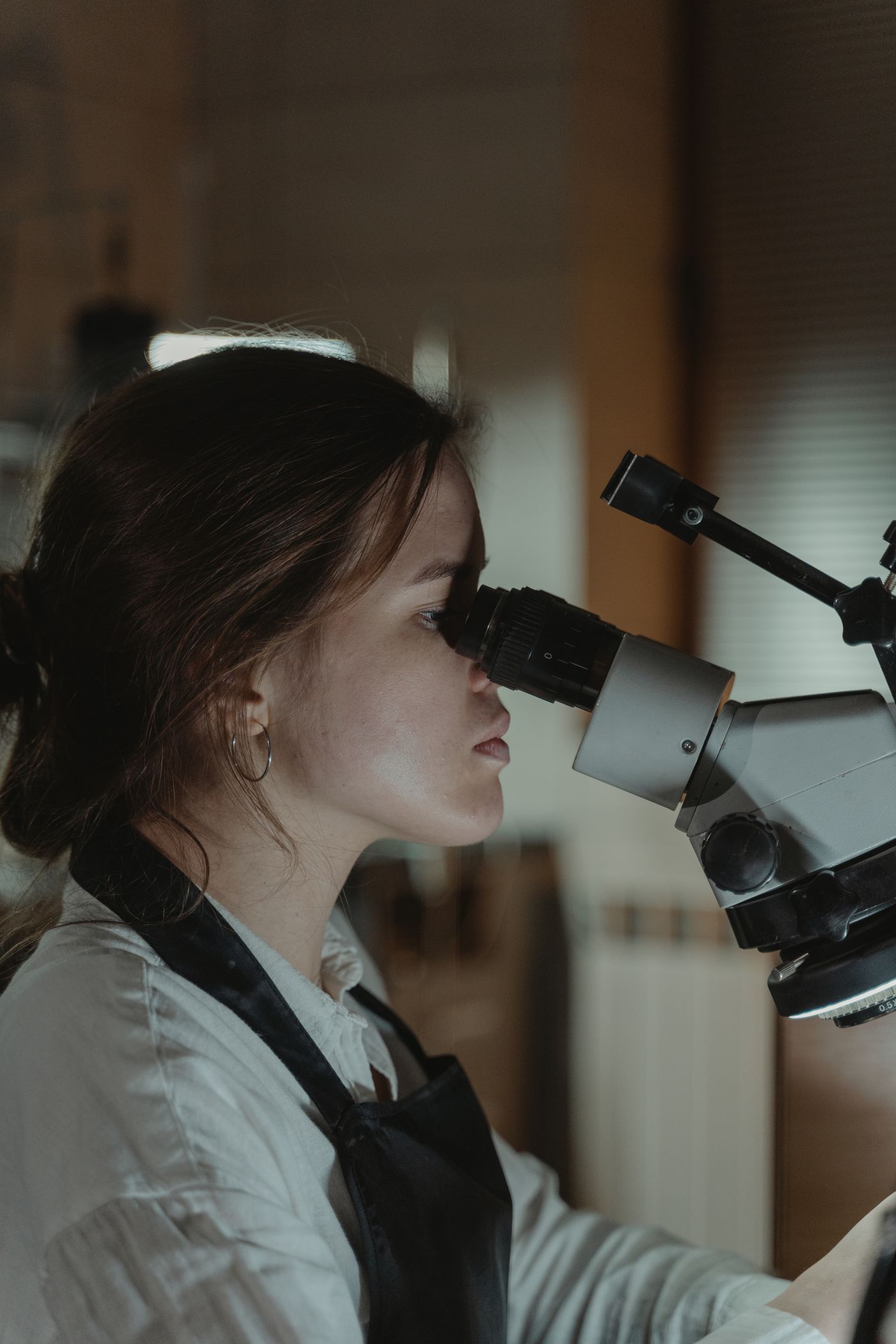
Lesson 02 — Fixation and the drying artefact
Why an air-dried Papanicolaou smear looks the way it does, how many seconds you actually have, and the side-by-side atlas fields that make the artefact obvious once seen. Ends with a preparation exercise on ten fields from the atlas.
§6
What is included
- 22 recorded lessons (streamed, 12 months)
- Downloadable image atlas — 400 annotated fields, permanent
- Staining troubleshooting chart (PDF)
- Screening exercises after every lesson
- Self-assessment with model answers
§7
Readership & prerequisites
Who it is written for
Cytotechnologists, residents and laboratory staff who prepare or screen cytological material.
What you need beforehand
None. Written to be the first structured cytology course for laboratory staff, and a refresher for residents.
§8
Assessment & certificate
40 screening questions from the atlas; pass mark 70 %. Two attempts included.
Passing the assessment adds this module to your MEDSMITH record. A certificate with hours studied is issued for the complete program; single modules receive a confirmation of completion on request.
§9
Delivery & access
How you receive it
After payment you land on your personal order page and receive access details by email within 24 hours on working days. Streamed lectures are watched in the browser on any device; downloadable material is yours permanently.
Several seats
Set the quantity in the cart; each seat receives its own access details. For a department of more than ten, see enrolment for departments.
Prices include VAT. The payment page may show the total in your local currency. Withdrawal terms for digital content are described in the access & withdrawal policy.
§10
Other modules
€490 €590
€590 €690
€690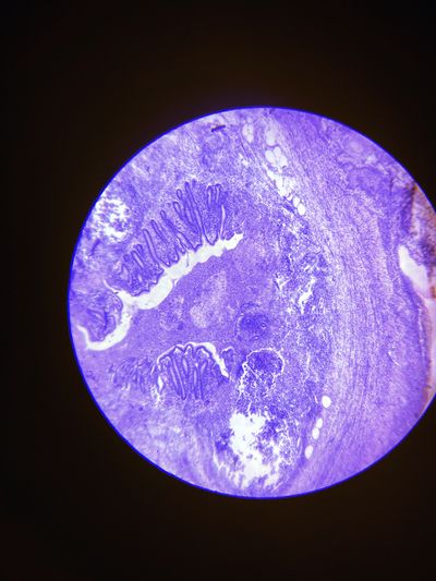 €1.190
 €1.690
 €2.990