Self-study web modules
9 h · 14 lessons
9 h · 14 lessons
€490VAT included
Details →
Seven self-study modules on digital slides, biopsy, cytology, gynaecological oncopathology and molecular diagnostics — written by pathologists who sign reports every week, for residents and laboratories that want to do the same with confidence.
MEDSMITH is a small medical research company in Bydgoszcz, Poland. Its founder is a doctor of medical sciences and pathomorphology specialist working in oncological diagnostics, with a European MSc in Molecular Pathology and research stays at Institut Curie and Radboud UMC. The company holds Polish patent rights for a biopsy needle guide and a device for preparing cytological smears — the same instruments that appear in the courses.
Recorded lecture series, a written reporting casebook and downloadable atlases, organised as seven numbered modules that can be taken alone or as one curriculum. Each module ends with self-assessment; the complete program issues a certificate of completion.
Everything is digital and self-paced. After payment you receive your personal access details by email within 24 hours. There are no live sessions, no fixed dates and nothing to ship — you study when the microscope is switched off.
Seven modules, numbered in the order we recommend — although each stands on its own. Prices include VAT; the payment page shows the total in your currency.
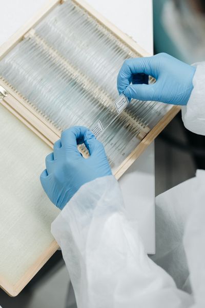
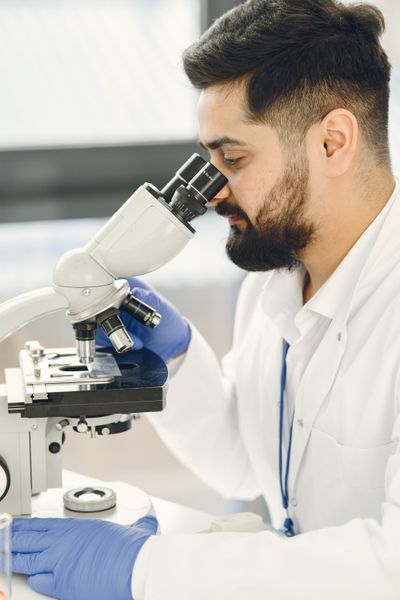
One table for the whole curriculum: what each module is made of, how long it takes and how long you keep it.
| No. | Module | Extent | Price |
|---|---|---|---|
| 01 | Foundations of Digital Pathology | 9 h · 14 lessons | €490 |
| 02 | Diagnostic Reporting Casebook | 120 cases · 6 chapters | €590 |
| 03 | Biopsy in Tumour Diagnosis | 11 h · 18 lectures | €690 |
| 04 | Cytopathology & Smear Preparation | 14 h · 22 lessons · 400 images | €890 |
| 05 | Ovarian & Gynaecological Oncopathology | 16 h · 24 lectures | €1.190 |
| 06 | Molecular Pathology in Diagnostic Practice | 18 h · 26 lectures | €1.690 |
| 07 | Diagnostic Oncopathology — Complete Program | 68 h · 104 lessons · 120 cases | €2.990 |
All prices include VAT and are per seat. Modules 01–06 bought separately come to €5.540; the complete program (07) contains all six.
Every recorded lesson has the same five parts, in the same order, so that after the second lesson you stop noticing the format and start noticing the slide.
Age, site, imaging, the question the clinician actually asked. Written on the screen exactly as it arrived in the laboratory.
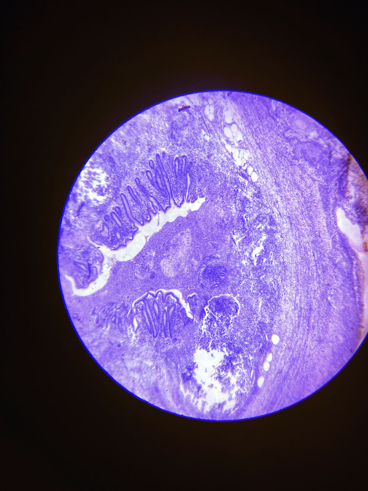
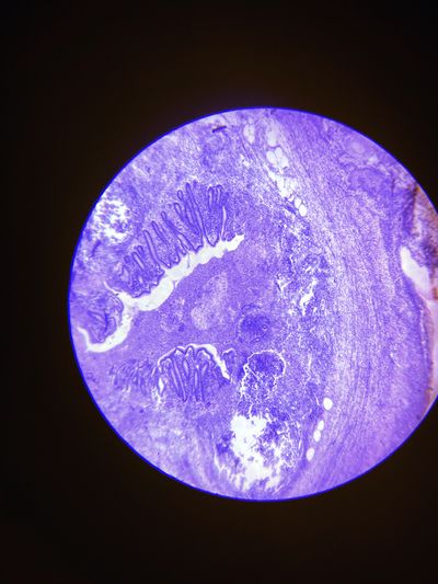
A whole-slide image or smear navigated live, from 2× architecture to 40× detail, with the author thinking aloud — including the fields that mislead.
Which two or three diagnoses are really in play and what immunostain, level or molecular test would separate them at the lowest cost in tissue.
The synoptic report as it was signed, with the sentence on uncertainty read out and explained.
Three to five new fields on the same theme, answered before the model answer appears. Results feed your module record.
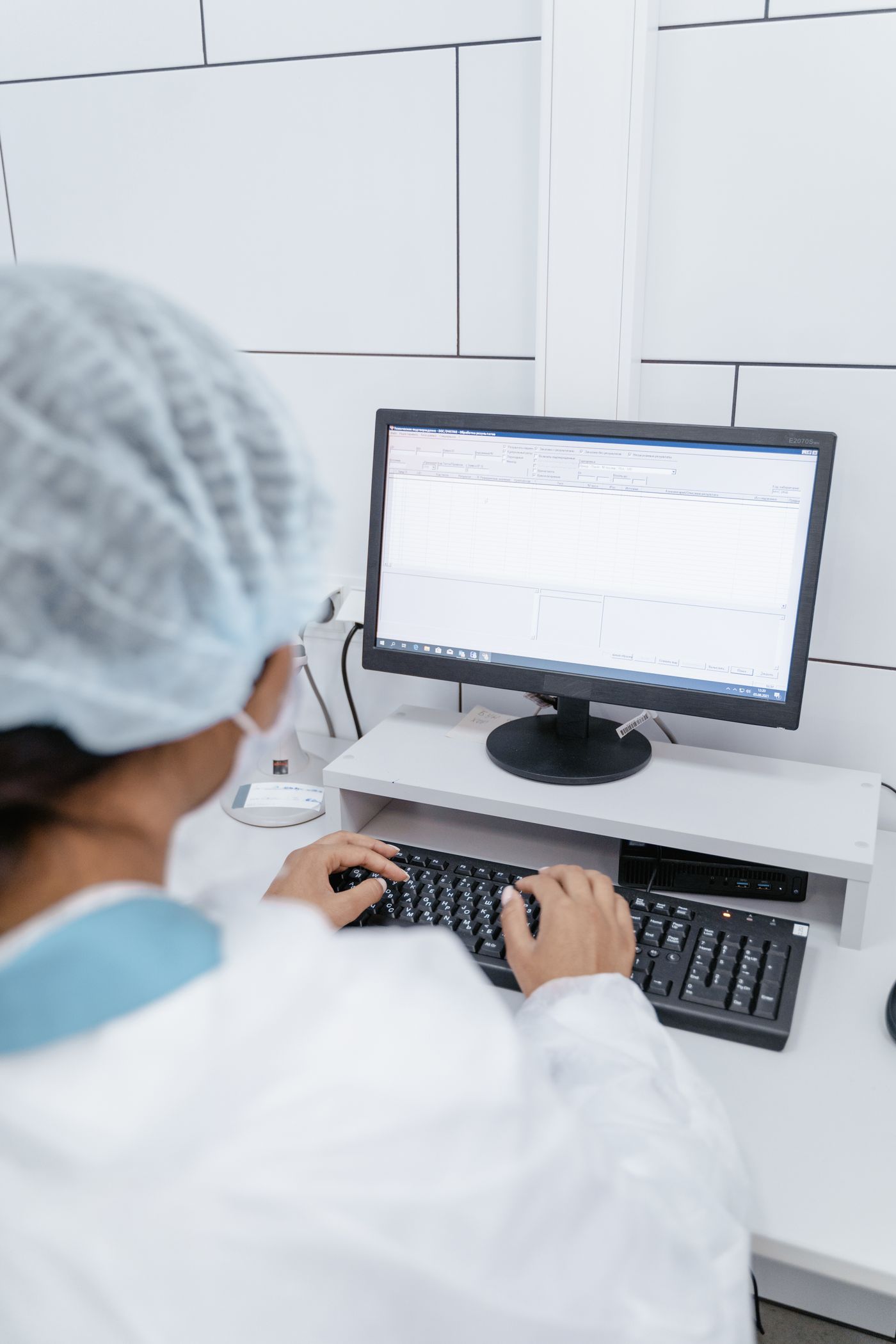
The modules follow the way a pathologist actually learns: look, describe, decide, report — then check against the answer.
Every lecture opens on a slide or a smear, not on a definition. Whole-slide images are shown at the magnifications you would use yourself.
Terminology follows WHO classifications and ICCR datasets, so what you learn transfers directly to your own synoptic reports.
Immunohistochemistry and molecular tests are introduced as questions you ask the slide, with cost and tissue budget in mind.
Self-assessment after each chapter with model answers. The complete program tracks progress and issues a certificate when all assessments are passed.
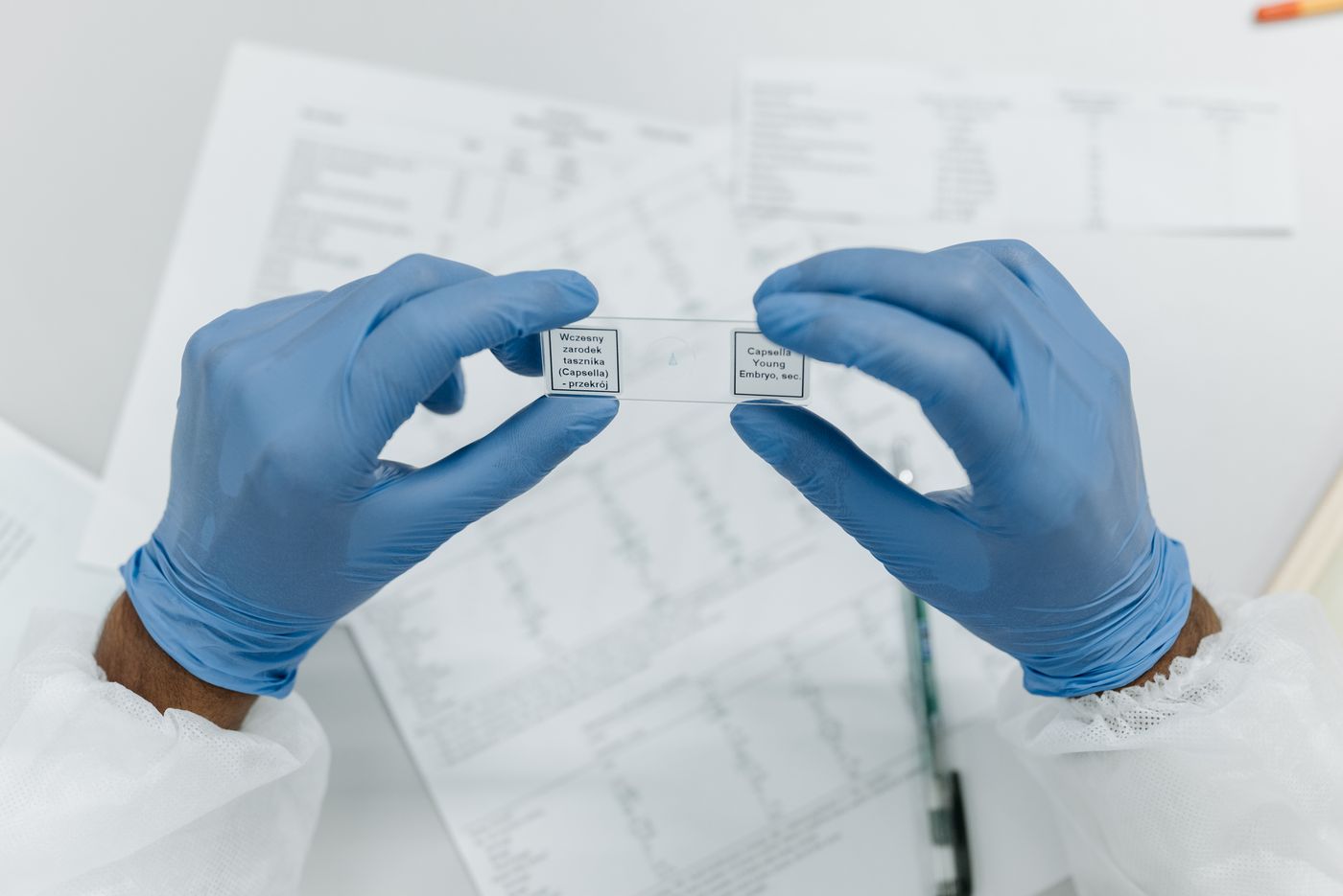
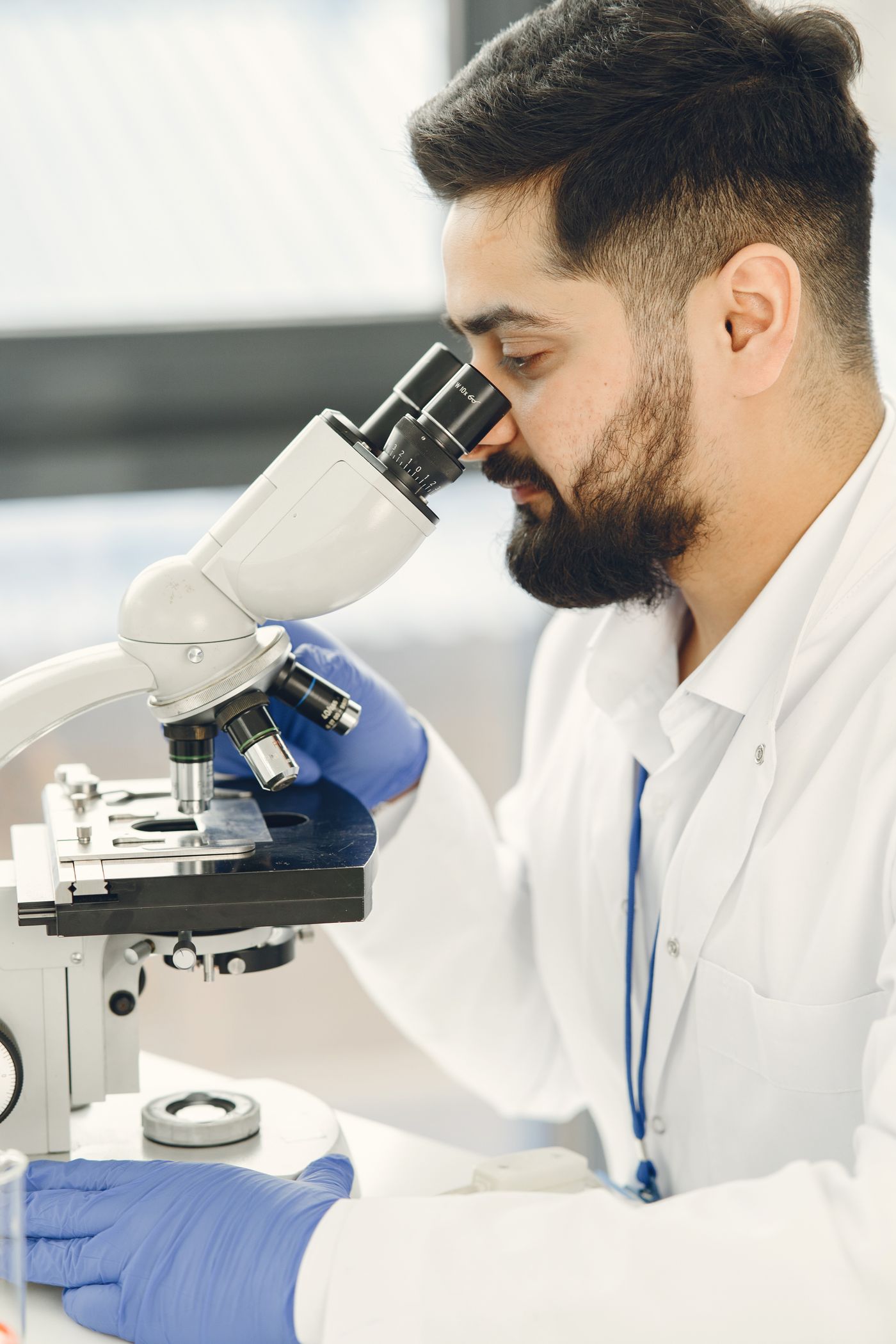
The curriculum is written and recorded by the company's founder, Adam Kowalewski, MD, PhD, and reviewed by colleagues in diagnostic and molecular pathology.
“A small biopsy is not a small diagnosis. The course exists because too much of what decides a report is never taught explicitly.”
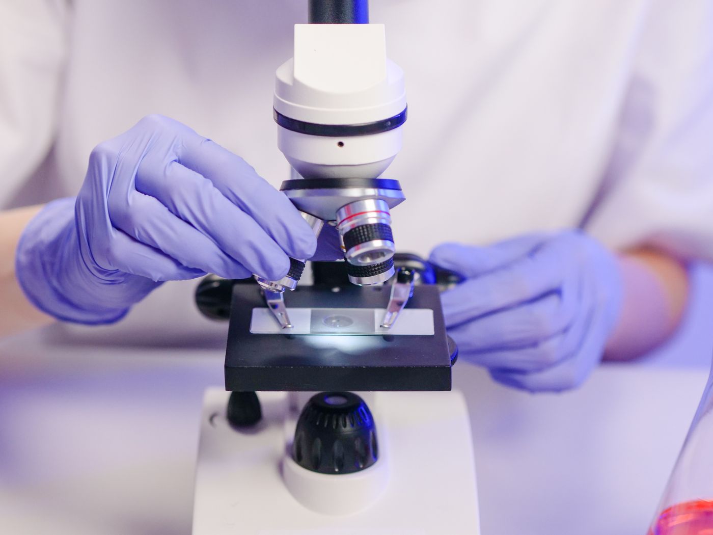
MEDSMITH began as a device company. Two instruments protected at the Polish Patent Office are the reason the biopsy and cytology modules exist — and appear in them as worked examples of pre-analytical quality.
Buy the one module you need this month, or the whole curriculum with longer access and a certificate. The arithmetic is below; both routes lead to the same lectures.
Diagnostic Oncopathology — Complete Program
Modules 01–06 · 68 hours of recorded and written study · integrated final paper passed
Issued to the name on the order, dated on the day the final paper is passed, signed by the author.
 MEDSMITH · Bydgoszcz · Vol. 1 · 2026
Assessment is there to tell you what you missed, not to gate the content. Every module closes with an image-based paper; the program adds one integrated paper across all six.
Twenty-six weeks at four to five hours a week takes a resident through the complete program. Access lasts 24 months, so the plan bends around on-call and examinations.
| Weeks 1–3 | Foundations of Digital Pathology — set up the viewer, learn the artefacts, run the validation exercise | 9 h |
| Weeks 1–26 | Diagnostic Reporting Casebook — two cases a week alongside everything else | 120 cases |
| Weeks 4–7 | Biopsy in Tumour Diagnosis | 11 h |
| Weeks 8–12 | Cytopathology & Smear Preparation, with the atlas screening exercises | 14 h |
| Weeks 13–18 | Ovarian & Gynaecological Oncopathology | 16 h |
| Weeks 19–25 | Molecular Pathology in Diagnostic Practice | 18 h |
| Week 26 | Integrated final paper; certificate issued on passing | 2 h |

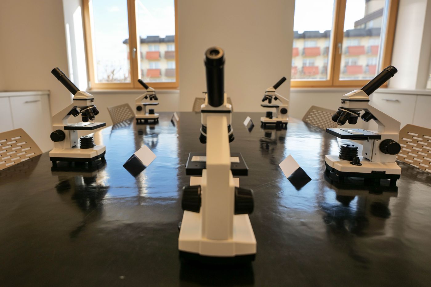
A structured route from digital slide basics to integrated molecular reporting, with a casebook for examination preparation.
Preparation technique that decides adequacy, screening systems and the artefacts that cost a diagnosis.
Oncologists, gynaecologists and interventional radiologists who want to understand what the report can and cannot say about their sample.

A department can enrol several trainees under one order and one payment. Seats are personal; progress is tracked per person and reported to the training lead on request.
Set the number of seats in the cart. Same price per seat, one payment, one order page.
Write to us for a departmental arrangement: staggered start dates, a named training lead, invoicing to the institution.
Access details for every seat are sent within one working day of payment, to the addresses you give us.
The modules teach against published classifications and reporting standards, not against the author's habits. These are the references the lessons cite most often.

Within 24 hours of payment (working days) you receive an email with your personal access details for the streamed lectures and download links for written material. Your order page, which opens right after payment, keeps a permanent record of what you bought and where to write.
Streamed modules are available for 12 months from the day access is granted (24 months for the complete program). Downloadable atlases, reference sheets and the casebook are yours permanently.
No. All modules are recorded or written and entirely self-paced. Questions about content can be sent by email; we answer within a few working days.
Yes — set the quantity in the cart. Each seat receives its own access details. For more than ten seats write to us for a group arrangement.
The displayed price includes VAT. The payment page may show the total in your local currency; our payment provider acts as merchant of record and issues the payment receipt. Cards, Apple Pay and Google Pay are accepted.
Until access is delivered you can cancel for a full refund. Because this is digital content, by asking for immediate access you agree that the statutory 14-day withdrawal right ends when access is delivered. See the access & withdrawal policy.
The complete program issues a MEDSMITH certificate of completion with the hours studied. Whether your national body accepts it towards continuing education depends on local rules — we are happy to provide the syllabus for your application.
Recorded material goes out of date. Lessons are re-recorded when a classification or guideline changes; everyone with active access receives the revised lesson at no charge, and the casebook's revised editions are sent for 24 months after purchase.
Found an error in a lesson or the casebook? Write to training@medsmith.pl — corrections are credited in the next revision.
Tell us your role and what you report most often. We reply by email within 24 hours on working days — training@medsmith.pl.
Write your question here. We read every message and reply by email within 24 hours on working days.